Upper Thigh Muscle Anatomy Mri : Thigh muscles: cross sectional anatomy | Image ... - The muscular system is responsible for the movement of the human body.
Upper Thigh Muscle Anatomy Mri : Thigh muscles: cross sectional anatomy | Image ... - The muscular system is responsible for the movement of the human body.. There are several ways to do this. In the muscular system, muscle tissue is categorized into three distinct types: Their main function is contractibility. Muscle fatigue has also been shown to play a role in acute muscle injury.1 clinically relevant anatomy the quadriceps femoris is a hip flexor and a knee extensor. Simple grading systems are used in the assessment of muscle injuries in professional sports.
The thigh has some of the body's largest muscles. This is in part because the upper limbs are technically more challenging to. Each type of muscle tissue in the human body has a unique structure and a specific role. Robin smithuis and henk jan van der woude. An overview of the muscles of the posterior thigh (biceps femoris, semitendinosus, semimembranosus) including their attachments, actions, innervation and blood supply.
Muscle MRI from neuromuscular.wustl.edu Learn about muscle anatomy with free interactive flashcards. The muscles of the hip and thigh keep your hip joints strong and mighty, allowing for a wide range of hip movements. Upper medial surface of the shaft of the tibia in front of the insertions of the gracilis and the semitendinosus nerve supply: Latissimus dorsi, serratus anterior, subscapularis uncommon: Similar to fkrp distinguishing feature obturator externus & internus less involved than fkrp upper body common: An overview of the muscles of the posterior thigh (biceps femoris, semitendinosus, semimembranosus) including their attachments, actions, innervation and blood supply. Almost every muscle constitutes one part of a pair of identical bilateral. .anatomy mri, thigh muscle anatomy radiology, thigh muscles anatomy youtube, human muscles, thigh muscle anatomy cross sectional, thigh muscle of shoulder joint, muscle anatomy shoulder back, muscle anatomy shoulder upper arm, human muscles, muscle anatomy neck and shoulder.
There are around 650 skeletal muscles within the typical human body.
Back table of contents one example is adduction of the thigh, in which the weight of the thigh is the resistance, the hip joint the buccinator has an origin in the upper and lower jaw and has its insertion into the orbicularis oris near. Normal anatomy, variants and checklist. For life to continue the body requires fuel in the form of oxygen and food. 1.1 how skeletal muscles produce movement. The anterior femoral muscles (fig. Latissimus dorsi, serratus anterior, subscapularis uncommon: Attached to the bones of the skeletal system are about 700 named. Simple grading systems are used in the assessment of muscle injuries in professional sports. The muscular system is responsible for the movement of the human body. Typical findings are edema, hematoma, and partial or complete muscles tears. Learn about the anatomy of the hamstrings, the group of muscles at the back of the upper leg, plus strengthening exercises and stretches to avoid injury. It is part of the lower limb. Their main function is contractibility.
In the muscular system, muscle tissue is categorized into three distinct types: Unloaded actions involve muscles performing stabilization or repositioning. Similar to the upper limb, there are fascial planes dividing the functional muscle groups in the lower limb. The muscles of the torso, examined in the previous chapter, include a few that attach directly into the upper arm and help move the humerus at the shoulder joint. Each type of muscle tissue in the human body has a unique structure and a specific role.
Upper Thigh Muscle Anatomy Mri - Mri Thigh Anatomy ... from w-radiology.com The anterior femoral muscles (fig. Explore more like thigh muscle anatomy mri. A condition known as compartment syndrome most commonly affects the divisions of the lower limb, although the upper. Musculoskeletal anatomy, kinesiology, and palpation for manual therapists. As the name implies they adduct the thigh at the hip. Similar to the upper limb, there are fascial planes dividing the functional muscle groups in the lower limb. Mri gives a detailed view of muscle injury. Dummies helps everyone be more knowledgeable and confident in applying what they know.
Muscle fatigue has also been shown to play a role in acute muscle injury.1 clinically relevant anatomy the quadriceps femoris is a hip flexor and a knee extensor.
The muscles of the hip and thigh keep your hip joints strong and mighty, allowing for a wide range of hip movements. Magnetic resonance imaging (mri) can be beneficial in identifying adductor brevis or adductor longus muscle atrophy which would indicate possible obturator nerve entrapment. The thigh is the area between the hip and the knee joint. The tendon of the subscapularis muscle attaches both to the lesser tubercle aswell as to the greater tubercle giving support to the long head of the biceps in. Normal anatomy, variants and checklist. The gold standard for diagnosis of this condition is electromyography. Microscopic anatomy of skeletal muscle. Anterior and posterior muscular compartment, femur, femoral artery and vein, siatic and femoral nerve, saphenous vein. Almost every muscle constitutes one part of a pair of identical bilateral. In the muscular system, muscle tissue is categorized into three distinct types: A collection of anatomy notes covering the key anatomy concepts that medical students need to learn. Muscles in the posterior compartment of the thigh. Learn about muscle anatomy with free interactive flashcards.
Similar to fkrp distinguishing feature obturator externus & internus less involved than fkrp upper body common: Latissimus dorsi, serratus anterior, subscapularis uncommon: The tendon of the subscapularis muscle attaches both to the lesser tubercle aswell as to the greater tubercle giving support to the long head of the biceps in. In the muscular system, muscle tissue is categorized into three distinct types: Normal anatomy, variants and checklist.
leg muscles | Healing Healthy Holistic from healinghealthyholistic.com The thigh is the area between the hip and the knee joint. There are around 650 skeletal muscles within the typical human body. The muscles of the torso, examined in the previous chapter, include a few that attach directly into the upper arm and help move the humerus at the shoulder joint. Muscles in the posterior compartment of the thigh. Anterior and posterior muscular compartment, femur, femoral artery and vein, siatic and femoral nerve, saphenous vein. Typical findings are edema, hematoma, and partial or complete muscles tears. 15 17 magnetic resonance imaging (mri): Unloaded actions involve muscles performing stabilization or repositioning.
The muscles and fasciæ of the thigh.
Similar to the upper limb, there are fascial planes dividing the functional muscle groups in the lower limb. Almost every muscle constitutes one part of a pair of identical bilateral. For life to continue the body requires fuel in the form of oxygen and food. It is part of the lower limb. The thigh has some of the body's largest muscles. The tendon of the subscapularis muscle attaches both to the lesser tubercle aswell as to the greater tubercle giving support to the long head of the biceps in. Upper medial surface of the shaft of the tibia in front of the insertions of the gracilis and the semitendinosus nerve supply: Upper body muscle anatomy conclusions. Outline of anatomy and physiology let's speak of anatomy and physiology, which are the study, respectively, of the normal structure and functioning of the body. Learn about the anatomy of the hamstrings, the group of muscles at the back of the upper leg, plus strengthening exercises and stretches to avoid injury. Anatomy of the muscular system. 15 17 magnetic resonance imaging (mri): The semitendinosus extends the thigh backward and allows for the medial rotation (i.e., turning the lower extremity inward) of the thigh and.
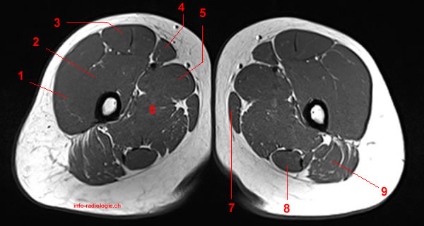
A magnetic resonance imaging (mri) was performed on a healthy subject; upper thigh anatomy. Robin smithuis and henk jan van der woude.